
Fig. 4.
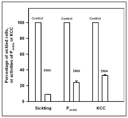
Effect of dimethyl adipimidate on sickling and K+ transport in Mg2+ clamped human sickle red cells. HbS cells samples were exposed to dimethyl adipimidate (6 mM) for 30 min at 37°C, pH 8, then washed three times to remove unreacted DMA; control samples were similarly treated but without DMA. Both were then placed in tonometers (40% haematocrit) and equilibrated with O2 at 150 mmHg or 100% N2. Cells were subsequently Mg2+ clamped, as described in the legend to Figure 1, except that only a single [Mg2+]o, 0.19 mM (plus 50μM EGTA), was used. Aliquots were removed, fixed in saline with 1% glutaraldehyde (pre-equilibrated with air or N2, as appropriate) for assessment of cell shape using light microscopy. Finally, K+ influxes were measured: Cl−-dependent K+ influx was taken as a measure of the activity of K+-Cl− cotransporter; the deoxygenation-induced Cl− independent K+ influx as a measure of Psickle activity. Ouabain (100 μM) and bumetanide (10 μM) were present. Data for K+ transport pathways or cell sickling are presented for cells in N2, normalised to 100% in the absence of DMA. Histograms represent means ± S.E.M. for 3 determinations.