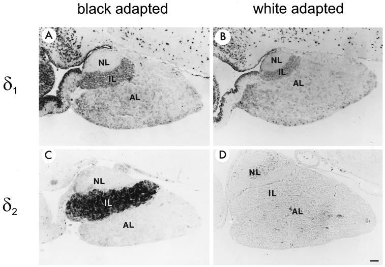
Figure 6.
Immunocytochemical analysis of δ1 and δ2 protein expression in the pituitary gland of X. laevis. Paraffin sections of pituitaries of either black- or white-adapted animals were incubated with affinity-purified anti-RH6 (1:50 dilution; A and B) or anti-1262N (1:1500 dilution; C and D) antibodies. NL, neural lobe. Bar, 100 μm.

