Figure 3.
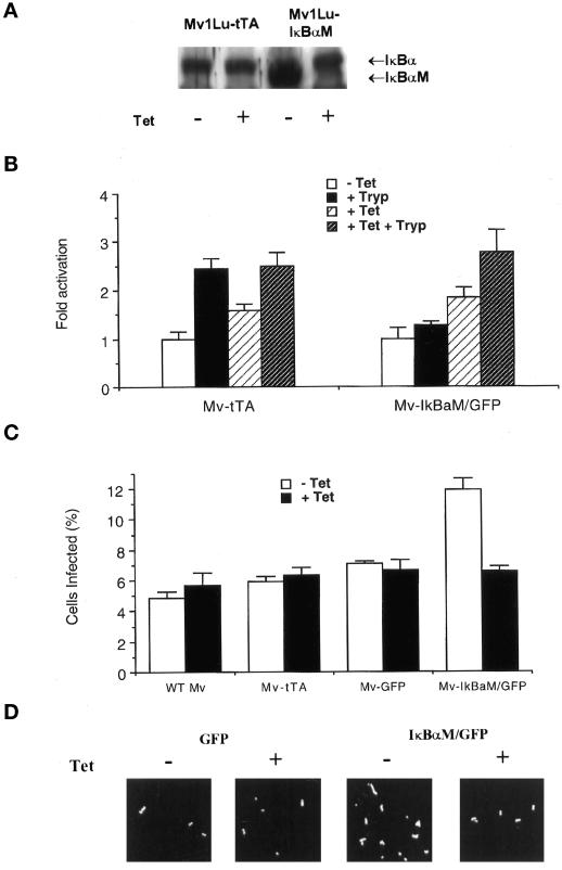
Expression of IκBaM enhances infection in epithelial cells. (A) Tetracycline-regulatable expression of IκBaM: Western blot of whole-cell lysates from tTA- or IκBaM-expressing Mv1Lu cells incubated overnight with or without 5 ng/ml tetracycline. Blots were incubated with anti-IκBa antibodies and visualized by ECL. Each lane contains 50 μg of protein. (B) Luciferase activity in pBXIIluc-transfected tTA- or IκBaM-expressing Mv1Lu cells (Mv) 24 h after exposure to RPMI and 1%BSA, Silvio strain trypomastigotes (1 × 10⁷/ml), tetracycline (+ Tet; 5 ng/ml), or tetracycline and trypomastigotes (+ Tet + Tryp; 1 × 10⁷/ml). Each point represents the mean of triplicate assays ± SEM. (C) Infection level in wild-type Mv1Lu (WT Mv) and in stable transfectants expressing tTA (Mv-tTA), GFP alone (Mv-GFP), or GFP with IκBaM (Mv-IκBaM/GFP) in the absence (− Tet) or presence (+ Tet) of 5 ng/ml tetracycline. Cells were incubated overnight with or without tetracycline before infection. Infections were stopped at 48 h. Each point represents the mean of triplicate assays ± SEM. At least 300 cells were counted per well. (D) Intracellular amastigotes in cells transfected with PTR5-DC/GFP or PTR5-IκBaM/GFP and infected with or without 5 ng/ml tetracycline as described above. Cells were fixed 48 h after infection, stained with human Chagasic sera, and visualized with TRITC-labeled anti-human immunoglobulin G (Sigma, St. Louis, MO). Similar numbers of host cells were present in each field.

