Figure 8.
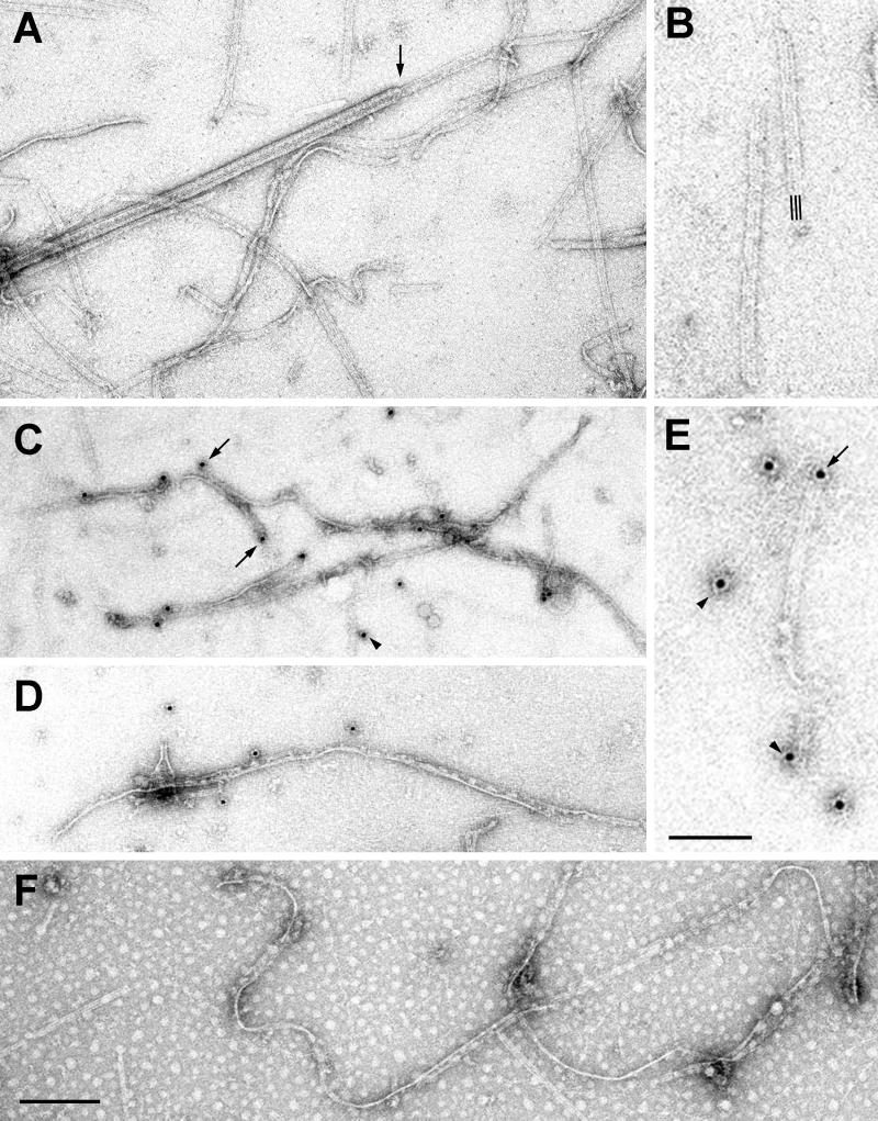
Negative stain and immuno-EM of Chlamydomonas ribbons. (A and B) Preparation of purified ribbons, negatively stained with 1% uranyl acetate. Ribbons are homogeneously composed of three protofilaments (three lines in B). Some ribbons are straight and rigid, and others are twisted and curled; some ribbons form pairs or sheets. A rare, contaminating A-microtubule is included for size comparison and to show the ribbon emerging from its end (arrow). (C–E) Ribbons stained with affinity-purified, rabbit anti-rib43aΔN64 antibodies, followed by 5-nm colloidal gold-conjugated goat anti-rabbit IgG. Ribbons are sparsely and randomly labeled along their length and at their ends (arrows). Many apparent background gold particles are actually due to label on short pieces of ribbons and on short, thin fibrils (arrowheads). (F) Control showing absence of staining when primary antibody is omitted. Bars, A, C, D, and F, 0.2 μm; B and E, 100 nm.

