Figure 9.
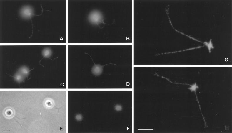
Immunofluorescence microscopy of whole cells and basal body–flagellar complexes. (A–D) Cells stained with affinity-purified rabbit anti-rib43aΔN64 antibody, followed by Texas Red-conjugated goat anti-rabbit IgG. (E) Phase-contrast image; (F) lack of staining with preimmune serum. (G and H) Immunofluorescence of isolated basal body–flagellar apparatuses stained with anti-rib43aΔN64. These results demonstrate that anti-rib43a antibodies specifically stain basal bodies and flagella in a punctate manner from base to tip. In addition, the anti-rib43a antibodies stained structures corresponding to the proximal portion of the four rootlet microtubules (G and H). Bars, A–F, 5 μm; G and H, 5 μm.

