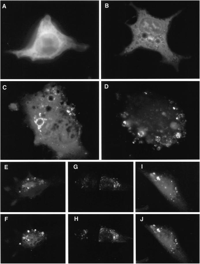
Figure 2.
Expression of GFP-tagged hVPS4 in cultured NRK cells. (A) Wild-type GFP-hVPS4 was transiently expressed in NRK cells and visualized by conventional fluorescence microscopy. (B) As in A, but with cells expressing GFP-hVPS4(KQ), an ATP binding-defective mutant. (C) As in A, but with cells expressing GFP-hVPS4(EQ), an ATP hydrolysis-defective mutant. (D) Cells expressing GFP-hVPS4(EQ) were treated with 0.01% (wt/vol) saponin immediately before fixation. NRK cells were cotransfected with GFP-hVPS4(EQ) and mSKD1-myc/His6(KQ) (E and F), hVPS4(EQ) and mSKD1-myc/His6(wt) (G and H), or hVPS4(KQ) and mSKD1-myc/His6(wt) (I and J). Cells were visualized by direct fluorescence microscopy (A–D, E, G, and I) or by indirect fluorescence microscopy using anti-myc antibody (F, H, and J).

