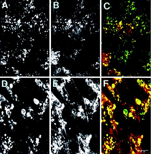
Figure 2.
The swollen lysosomal vacuoles of GlcNAc-TV transfectants are labeled with L-PHA. Untransfected Mv1Lu (A–C) or GlcNAcTV-transfected M9 (D–F) cells plated for 6 d were double immunofluorescently labeled with anti-LAMP-2 followed by FITC-conjugated anti-mouse secondary antibody (A and D) or with rhodamine-conjugated L-PHA (B and E). Merged images are presented in C and F (LAMP-2 in green, L-PHA in red). LAMP-2- and L-PHA-reactive β1–6-branched oligosaccharides are localized to lysosomes of Mv1Lu cells as well as to the large lysosomal vacuoles of GlcNAc-TV M9 transfectants. Bar, 10 μm.

