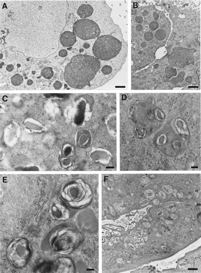
Figure 5.
Formation of MLBs via autophagic vacuole degradation after leupeptin washout. GlcNAc-TV-transfected M1 cells were plated for 2 d and then incubated for 4 d in the presence of 2 μg/ml leupeptin (A) and then washed and incubated in leupeptin-free medium for 15 (B), 24 (C), 48 (D and E), or 72 (F) h before being processed for electron microscopy. After leupeptin treatment, MLBs disappear, and large autophagic vacuoles are present (A). After 24 and 48 h, single or multiple foci of lamella form within the autophagic vacuole (C–E), and after 72 h the autophagic vacuoles transform into lamellar structures resembling those of untreated cells (F). Bars, 1 μm (A, B, and F); 0.2 μm (C–E).

