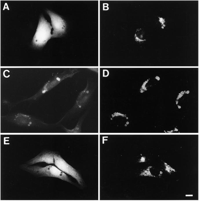
Figure 8.
Incorporation of cytosolic FITC-dextran into MLBs is inhibited by 3-MA. M9 cells were scraped from plastic dishes in the presence of 2.5 mg/ml FITC-dextran, washed extensively, replated on coverslips in regular medium (A–D) or medium containing 10 mM 3-MA (E and F), and then fixed after 2 (A and B) or 48 (C–F) h and labeled for LAMP-2 using Texas Red-conjugated secondary antibodies. The distribution of FITC-dextran (A, C, and E) and LAMP-2 (B, D, and F) in the same cells is presented. After only 2 h of plating, FITC-dextran is cytosolic and excluded form large LAMP-2-positive vacuoles (A and B); however, with time FITC-dextran is incorporated via autophagy into LAMP-2-positive perinuclear vacuoles equivalent to MLBs (C and D). Autophagic incorporation of FITC-dextran into MLBs is inhibited by 3-MA (E and F). Bar, 10 μm.

