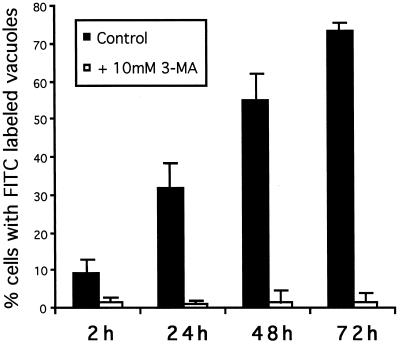
Figure 9.
Autophagy of scrape-loaded FITC-dextran in GlcNAc-TV-transfected cells. FITC-dextran was scrape loaded into M9 cells as in Figure 8 and incubated in regular medium (filled bars) or medium supplemented with 10 mM 3-MA (open bars) for 2, 24, 48, or 72 h (as indicated) before fixation and labeling for LAMP-2 as in Figure 8. Fifty FITC-dextran-loaded cells per slide were assessed for the presence of FITC labeling in LAMP-2-positive vacuoles. The percent of cells that exhibit FITC labeling of lysosomal vacuoles is presented and represents the average ± SD of six experiments.

