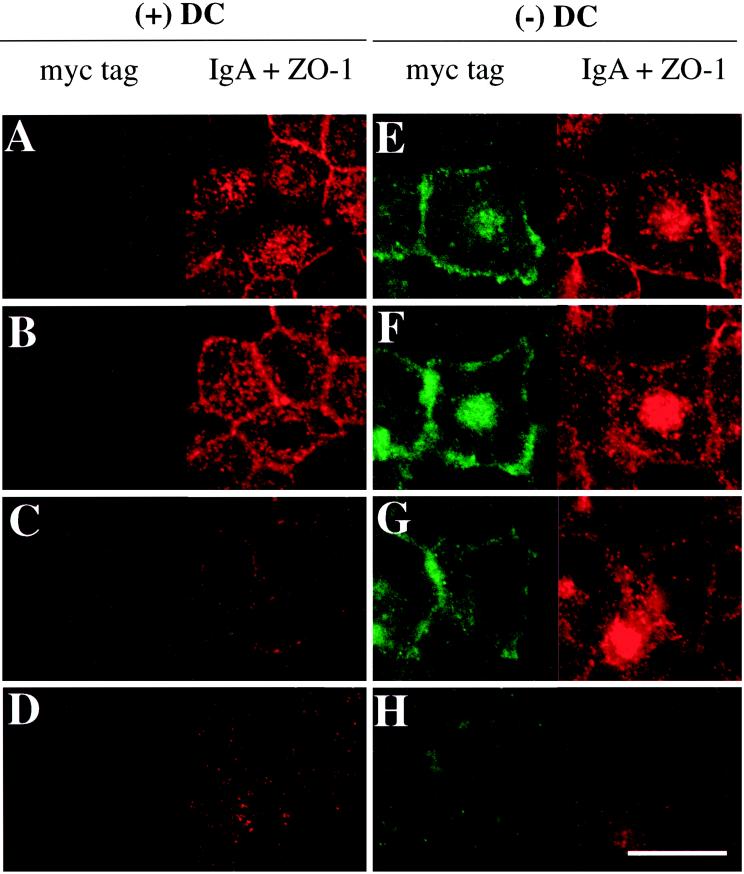
Figure 2.
Distribution of basolaterally internalized IgA and myc-tagged Rac1V12 in cells grown in the absence (−) or presence (+) of DC. IgA was internalized from the basolateral surface for 10 min at 37°C in Rac1V12 cells grown in the presence (A–D) or absence (E–H) of DC and then washed and chased for 5 min at 37°C. Cells were fixed with paraformaldehyde and stained with the appropriate antibodies, and FITC and Cy5 emissions (which are displayed in the left and right halves of each panel, respectively) were captured with the use of a scanning laser confocal microscope. Shown are optical sections from the base of the cells (D and H), along the lateral surface of the cells (C and G), above the nucleus (B and F), and at or above the level of the tight junctions (A and E). Note that there are at least 2–3 μm between each optical section. The tight junctions are the thin red lines that surround the cell. Bar, 10 μm.

