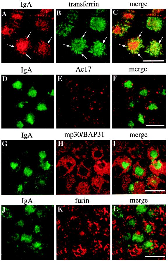
Figure 3.
Distribution of IgA, Tf, the Ac17 antigen, mp30/BAP31, and furin in cells expressing Rac1V12. IgA was internalized from the basolateral surface of the cell for 10 min at 37°C, washed, and then chased for 60 min at 37°C. (A–C) Tf was internalized during the last 10 min of the 60-min chase. The cells were fixed, incubated with antibodies against IgA and Tf (A–C), IgA and Ac17 antigen (D–F), IgA and mp30/BAP31 (G–I), or IgA and furin (J–L) and then reacted with the appropriate secondary antibody coupled to FITC or Cy5. Arrows indicate regions of colocalization. A single optical section at the level of the central aggregate was obtained with a scanning laser confocal microscope. Bar, 10 μm.

