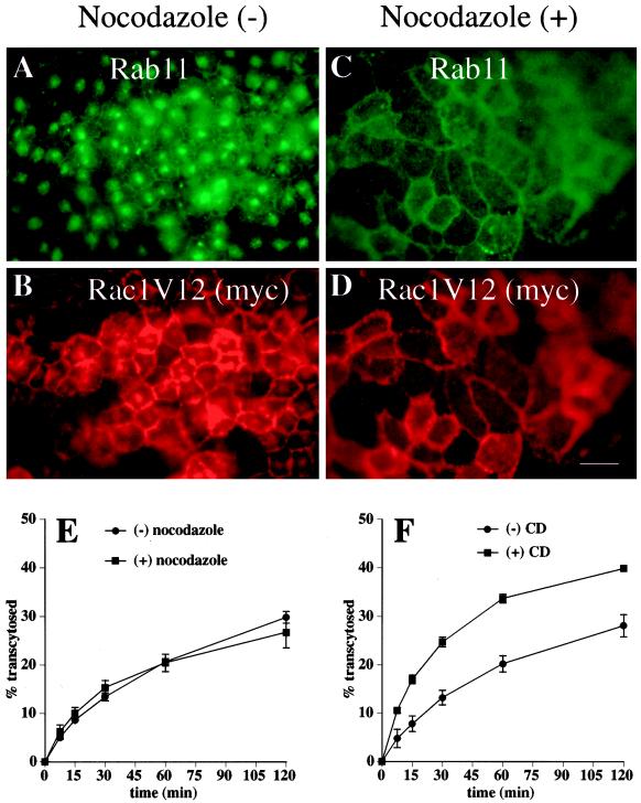
Figure 8.
Effects of nocodazole and CD on the distribution and exit of proteins from the central aggregate. (A–D) Cells were mock treated or treated with nocodazole, fixed, incubated with antibodies against rab11 and the myc tag, and then reacted with the appropriate secondary antibody coupled to FITC or Texas red. Bar, 10 μm. (E) [125I]IgA was internalized from the basolateral surface of the cells for 10 min at 37°C, and the cells were washed and then chased for 60 min. Cells were rapidly chilled to 4°C for 60 min (with [+] or without [−] nocodazole), and the postendocytic fate of ligand was assessed in a subsequent 120-min incubation at 37°C (with or without nocodazole). The percentage of total ligand released apically (transcytosed) is shown. Values for degradation were as follows: without nocodazole, 21.0 ± 1.2%; with nocodazole, 21.9 ± 0.8%. Values for ligand remaining cell associated were as follows: without nocodazole, 36.1 ± 5.5%; with nocodazole, 37.2 ± 1.5%. (F) [125I]IgA was internalized from the basolateral surface of the cells for 10 min at 37°C, and the cells were washed and then chased for 45 min at 37°C. After a 15-min treatment with (+) or without (−) 25 μg/ml CD, the postendocytic fate of ligand was assessed in a 120-min incubation at 37°C (with or without CD). The percentage of total ligand released apically (transcytosed) is shown. Values for degradation were as follows: without CD, 10.2 ± 0.5%; with CD, 14.0 ± 1.1%. Values for ligand remaining cell associated were as follows: without CD, 46.7 ± 2.2%; with CD, 35.7 ± 1.1%. Values (mean ± SD; n = 3) are from a representative experiment.

