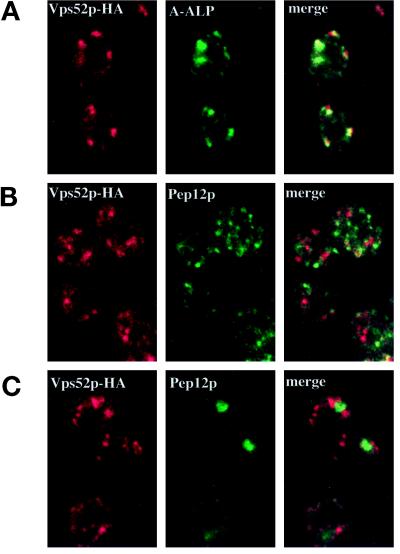
Figure 10.
Vps52p-HA colocalizes with A-ALP at the late Golgi by indirect immunofluorescence microscopy. (A) Vps52p-HA–expressing cells (LCY226) containing pSN55 (A-ALP) were fixed, spheroplasted, and double labeled with an anti-HA polyclonal antiserum and an anti-ALP mAb. Pairs of images corresponding to each antigen were collected with the use of confocal microscopy, and the images were merged to show coincidence of the two staining patterns. A majority of the structures that labeled with the HA antibody also contained A-ALP, although the relative intensities of the two staining patterns varied. (B) LCY226 (Vps52p-HA) cells and (C) LCY243 (Vps52p-HA vps27Δ) cells double labeled with an anti-HA polyclonal antiserum and a mAb to Pep12p.

