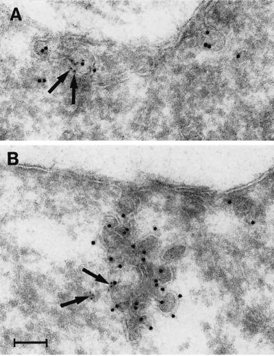
Figure 10.
Localization of caveolin-1 and filamin in CNF-1–stimulated (24 h) T4.5 trophoblasts with the use of immunoelectron microscopy. (A) A small cluster of two caveolae is positive for caveolin-1 (10-nm gold). Filamin (5-nm gold; arrows) is present at the caveolar membrane. (B) Large racemose cluster of caveolae labeled for caveolin-1 (10-nm gold) and filamin (5-nm gold; arrows). The caveolae cluster invaginates deeply into the cell but is still surface connected. Bar, 100 nm.

