Figure 7.
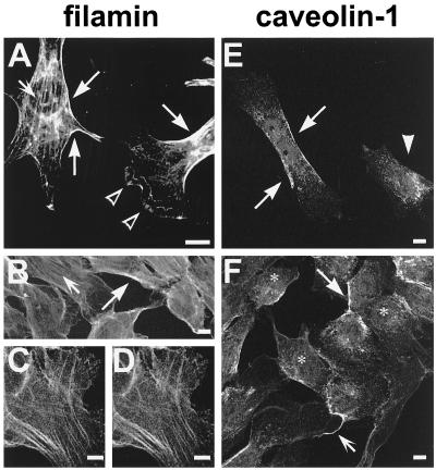
Confocal images of NIH/3T3 fibroblasts (A) and T4.5 trophoblasts (B–F) stained for filamin (A–D) or caveolin-1 (E and F). Filamin immunoreactivity was dominant on stress fibers (A and B, small arrows) and at the cell periphery (A and B, large arrows). Blurry immunoreactivity was seen in lamellipodia (A, open arrowheads). The labeling produced by antibodies mab1680 and pab228 is identical (compare C and D). Caveolin-1 was detected in patches or in a punctate pattern at the plasma membrane (E and F, large arrows) and along cellular processes (F, small arrow). The arrowhead in E designates a cell apparently showing only intracellular caveolin-1 immunoreactivity. Antibodies used were pab228 (A and C), mab1680 (B and D), and polyclonal anti-caveolin-1 (E and F). Bars, 10 μm.

