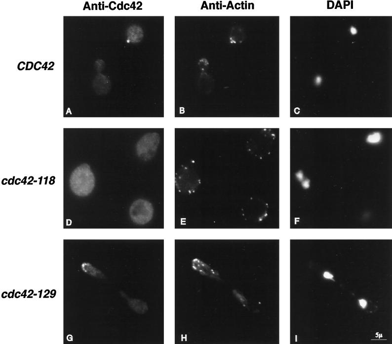
Figure 6.
Fluorescence images of wild-type (CDC42; DDY1300; top row), cdc42-118D76A (DDY1326), and cdc42-129V36T (DDY1344) cells shifted from 25 to 37°C for 6 h and probed with affinity-purified Cdc42 peptide antibody (left column), affinity-purified anti-actin antibody (middle column), or DAPI for DNA (right column).

