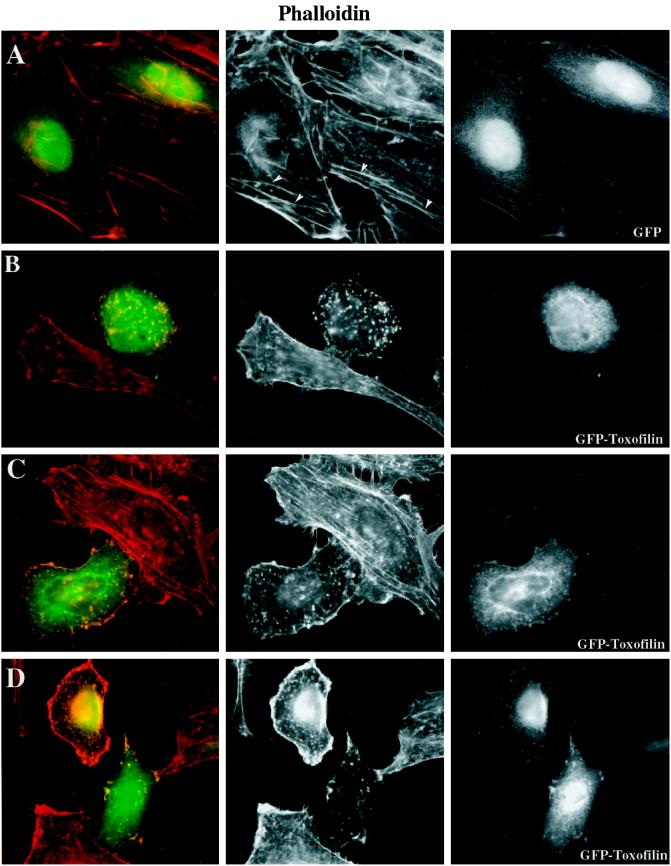
Figure 4.
Expression of GFP-Toxofilin fusion protein affects actin dynamics in mammalian nonmuscle cells. HeLa cells (50% confluent) were transfected by the CaCl2 phosphate method with plamids encoding either GFP-Toxofilin or GFP (see MATERIALS AND METHODS) and were incubated overnight before replating on glass coverslips. The cells were incubated for 20 h (37°C, 5% CO2) before processing for immunofluorescence (see MATERIALS AND METHODS). For revealing the F-actin or Toxofilin, the cells were permeabilized after fixation and stained with either rhodamine-phalloidin or with an anti-Toxofilin antibody, respectively. (A) Phalloidin staining of F-actin in HeLa cells expressing GFP. Well-organized actin stress fibers are visible. (B–D) Phalloidin staining of F-actin in cells expressing GFP-Toxofilin. Actin stress fibers are disorganized. (D) Different levels of expression of the GFP-Toxofilin. (E) Detection of Toxofilin with an anti-Toxofilin antibody in HeLa cells transfected with the GFP-Toxofilin-encoding plasmid.


