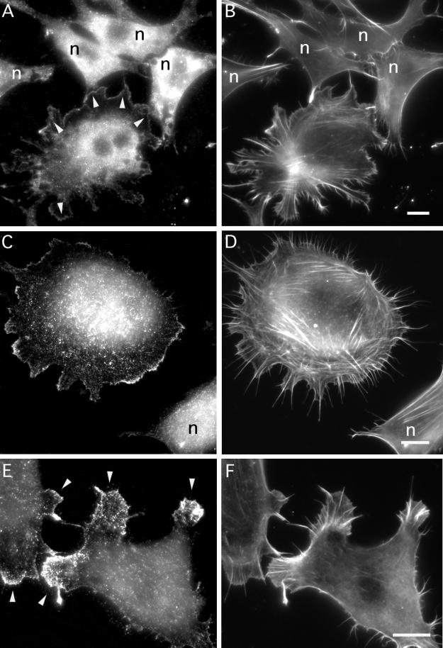
Figure 10.
Evidence that translocation of mAbp1 to the cell periphery is Rac1 mediated but Src independent. Serum-starved NIH 3T3 cells were transfected with mycRac1L61. (A and B) Field of nontransfected cells (marked by n throughout) and one Rac1L61-positive cell, which shows lamellipodia formation and mAbp1 translocation (some such areas are marked by arrowheads in A) despite prolonged serum starvation. (C–F) Examples of Rac1L61-transfected cells displaying accumulation of mAbp1 at the leading edge of lamellipodial areas (labeled by arrowheads in E) at higher magnifications. (A, C, and E) mAbp1 immunolabeling; (B, D, and F) F-actin staining with rhodamine-phalloidin. Bars, 10 μm.

