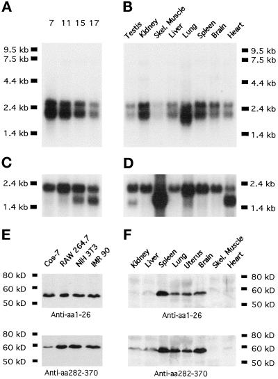
Figure 3.
Mammalian Abp1 mRNA and protein expression levels. (A) mAbp1 mRNA expression examined by Northern blotting during the course of mouse embryo development; the numbers on top of each lane represent days after fertilization. (B) Mouse Abp1 mRNA levels in different adult tissues. The Northern probe comprised the proline-rich domain (303 nucleotides corresponding to aa 282–382 of mAbp1). (C and D) Actin expression is shown as a control for RNA integrity and transfer efficiency. (E and F) Mammalian Abp1 protein on immunoblots is detected by both antibody GP1 (anti-N terminus) and antibody GP5 (anti-proline-rich domain) as a single band of 56 kDa. (E) Four representative cell lines are shown; 50 μg of protein from postnuclear cell homogenates were loaded per lane. (F) mAbp1 levels in different tissues from adult mice; 250 μg of protein were loaded per lane; both blots were incubated and developed together.

