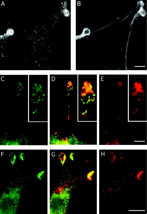
Figure 4.
Indirect immunolabelling of endogenous mAbp1 reveals a punctate distribution colocalizing with the cortical actin cytoskeleton at sites of cellular growth. (A, C, D, G, and H) Indirect immunolabeling of mouse Abp1. F-actin staining by rhodamine-phalloidin is shown in B, D, and E. Rab8 labeling of exocytic vesicles is shown in F and G. (A and B) NIH 3T3 cells. Bar, 10 μm. (C–E) Protrusion of a fetal human IMR90 fibroblast; the insets show a 2.25× higher magnification of the protrusion tip. Abp1 immunolocalization is shown in green (C), and F-actin stained with rhodamine-phalloidin is in red (E); areas of colocalization appear as different shades of yellow in the merged image (D). Bar, 10 μm. (F–H) Rab8 labeling (green; F), Abp1 labeling (red; H) in NIH 3T3 fibroblasts; areas of colocalization appear as different shades of yellow in the merged image (G). Bar, 10 μm.

