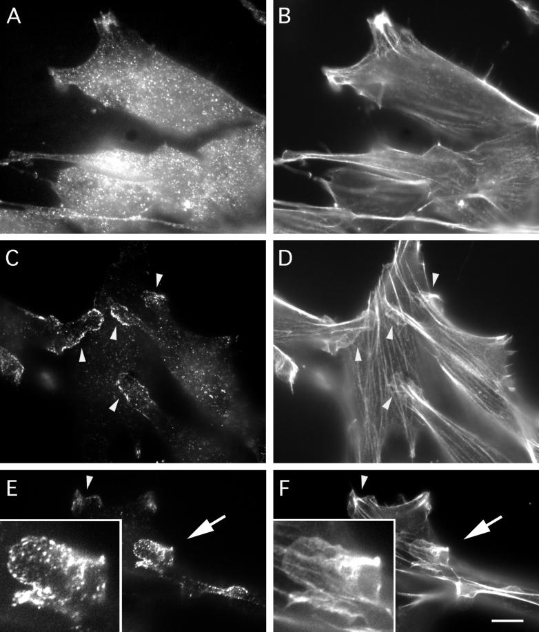
Figure 5.
mAbp1 localization in extracted and unextracted NIH 3T3 cells. (A and B) Immunolocalization of mAbp1 (A) and rhodamine-phalloidin localization of F-actin (B) in cells fixed before extraction. (C–F) Immunolocalization of mAbp1 (C and E) and rhodamine-phalloidin localization of F-actin (D and F) in cells briefly extracted with Triton X-100 before fixation. Arrowheads identify examples of areas showing association of mAbp1 with lamellipodial actin. Insets in E and F are 2.5× enlargements of a large lamellipodium (arrow) at the end of a cell protrusion that appears to be crawling over another cell. Bar, 10 μm.

