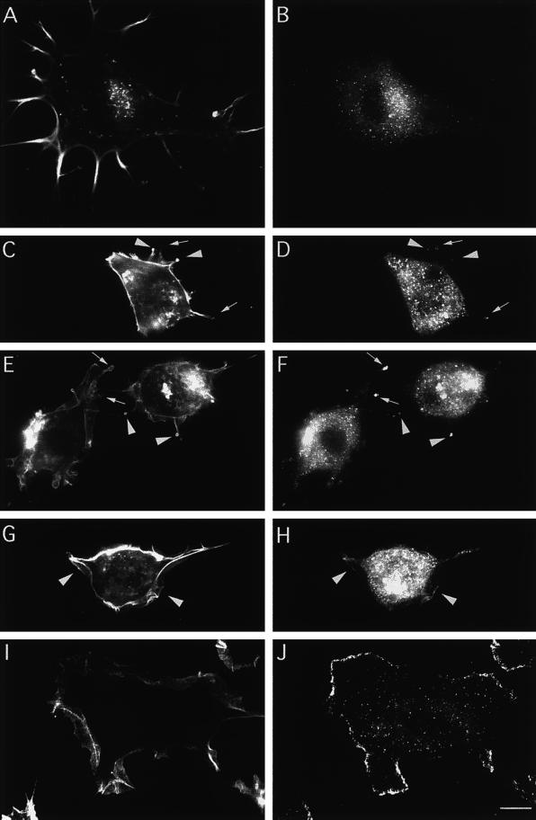
Figure 8.
Abp1 distribution is rapidly shifted toward the periphery of NIH 3T3 cells in response to growth factors that lead to Rac1 activation (here addition of 5 ng/ml PDGF). F-actin was stained with rhodamine-phalloidin (A, C, E, G, and I), and mAbp1 was detected by anti-mAbp1 antibody GP5 and FITC-conjugated secondary antibody (B, D, F, H, and J). (A and B) Quiescent cells starved for serum overnight. (C–J) Different time points of the response to growth factor: 1 min (C and D), 2 min (E and F), 4 min (G and H), and 10 min after PDGF addition (I and J). Arrowheads indicate protrusive areas with high F-actin content (C and E) at early time points at which mAbp1 was detected and accumulated (D and F). Arrows in C–F identify areas characterized by high mAbp1 accumulation with lower cortical actin accumulation. Arrowheads in G and H mark areas where the first continuous leading edge mAbp1 labeling at newly forming lamellipodia occurred at 4 min. Bars, 10 μm.

