
Figure 1.
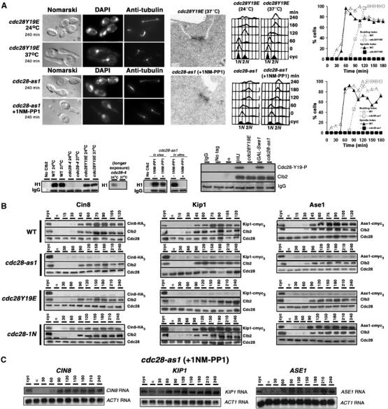
Low endogenous levels of microtubule-associated proteins in cdc28–as1 and cdc28Y19E cells. (A) cdc28Y19E (top panel) and cdc28-as1 (middle panel) cells were arrested with α factor and released at 37°C and 24°C in presence of 1NM-PP1, respectively. Electron micrographs show state of SPBs at 240 min. Plots show spindle and budding index. (Lower panel) Western blots show histone H1 kinase activity and tyrosine phosphorylation status. (B) Wild-type (WT), cdc28-as1, cdc28-Y19E and cdc28-1N cells carrying endogenously tagged Cin8-HA3, Kip1-cmyc3 or Ase1-cmyc3 were synchronized in G1 with α factor treatment and then released into at 24°C in medium containing 1NM-PP1 (for cdc28-as1) or 37°C (for cdc28Y19E and cdc28-1N). Samples for Western blotting were taken at 15-min intervals for WT and 30-min intervals for cdc28 mutants for comparison (doubling time in cdc28 mutants is approximately twice that of WT). (C) G1-synchronised cdc28-as1 cells were released in the presence of 1NM-PP1. Samples were collected at 30-min intervals for RNA isolation and subsequent Northern blotting.