
Figure 4.
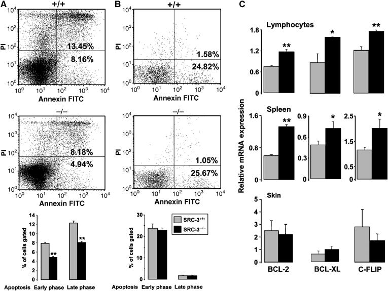
Decreased apoptotic activity in SRC-3−/− mice. Flow cytometric analysis of apoptotic SRC-3+/+ and SRC-3−/− spleen cell suspensions (A) and MEFs (B) after staining by annexin V FITC and propidium iodide (PI). The bar graphs at the bottom of (A) and (B) show the quantification of three independent experiments **P<0.01; (C) mRNA levels of BCL-2, BCL-XL, and c-FLIP in lymphocytes, spleen, and skin of SRC-3+/+ and SRC-3−/− animals (age 7 weeks, n=8) determined by quantitative RT–PCR. *P<0.05, **P<0.01.