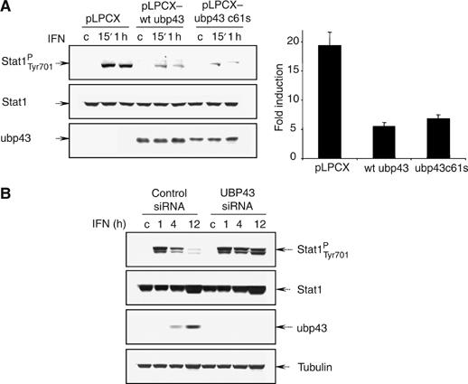
Figure 3.

Ectopic expression of Ubp43 blocks STAT1 phosphorylation and IFN-mediated gene induction. (A) STAT1-deficient U3A stable cell lines expressing vector-control, wt mUbp43, or mUbp43C61S were transiently co-transfected by STAT1 and ISRE-driven luciferase reporter plasmid. At 24 h post-transfection, cells were either left untreated or treated with hIFN-α for 15′, 1 h, and 16 h. Level of STAT1 phosphorylation and expression was assessed by Western blotting with the respective antibodies (left). Luciferase activities were measured, normalized, and presented as fold increase of relative luciferase activity in IFN treated cells (at 16 h point) over the untreated controls (right). The error bars indicate the s.d. of the mean. (B) KT-1 cells were stably transfected with control siRNA, hUBP43-specific siRNA. After 1 week of drug selection, cells were either left untreated or treated with hIFN-α for 1, 4 or 12 h respectively. STAT1 phosphorylation and expression was assessed by Western blotting with the respective antibodies. Specific inhibition of endogenous hUBP43 by siRNA in the respective stable lines was confirmed by Western blotting with anti-hUBP43-specific antibodies.
