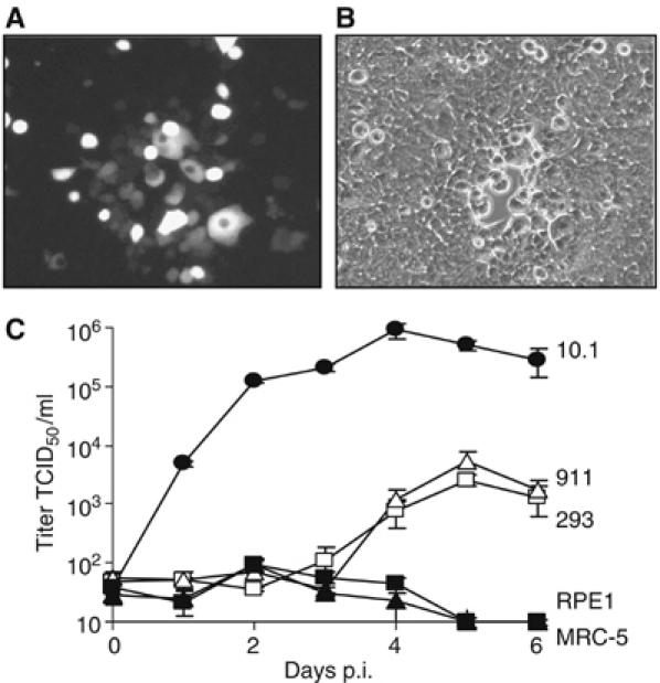
Figure 1.

MCMV replication in human 293 and 911 cells. (A, B) Fluorescent and phase contrast images of 293 cells 6 days after infection with MCMV-GFP at a low MOI. (C) Growth kinetic of MCMV-GFP on murine 10.1 cells and human 293, 911, RPE1 and MRC-5 cells. Cells were infected at an MOI of 5 TCID50/cell. Virus titers were determined in the supernatant.
