
Figure 7.
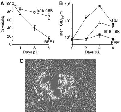
Growth of rat cytomegalovirus in human cells. (A) RPE1 cells died after infection with RCMV at an MOI of 5 TCID50/cell, but RPE1 cells expressing E1B-19k were mostly protected from RCMV-induced cell death. (B) RPE1 cells, RPE1-E1B-19k cells, and rat embryo fibroblasts (REF) were infected with RCMV at an MOI of 5 TCID50/cell, and titers in the supernatant were determined. (C) Phase contrast image of RPE1 cells expressing E1B-19k 7 days after low-MOI infection with RCMV. Plaque formation as an indication of virus replication and spread was only observed in RPE1-E1B-19k cells, but not in normal RPE1 cells.