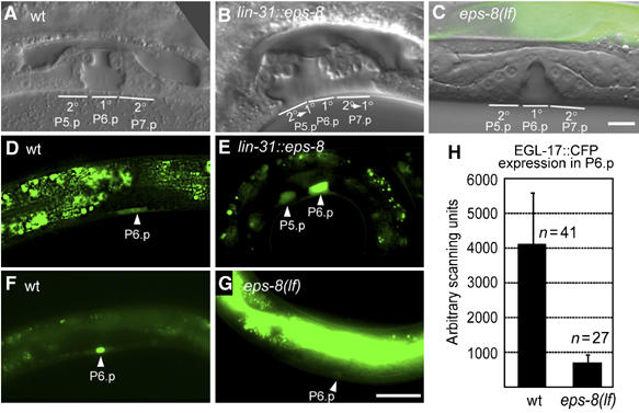
Figure 1.

EPS-8 positively regulates vulval cell fate specification. (A) Morphology of a wild-type vulva at the L4 stage. The position of the 2° descendants of P5.p and P7.p and the 1° descendants of P6.p (out of focus) is indicated. Note that some of the 2° vulval cells remain attached to the cuticula while all 1° cells are detached. (B) Vulval morphology in a lin-31::eps-8a L4 larva showing a 2° towards 1° fate transformation of P5.p and P7.p descendants. (C) eps-8(lf) L4 larva with a wild-type vulva consisting of 22 cells. (D) Expression of the 1° cell fate marker EGL-17::GFP in a wild-type early L3 larva is restricted to P6.p. (E) Ectopic EGL-17::GFP expression in P5.p in a lin-31::eps-8a L3 larva (17% of the cases, n=24). (F) EGL-17::CFP expression in the nucleus of P6.p in a wild-type L3 larva. (G) Reduced EGL-17::CFP expression in P6.p in an eps-8(lf) L3 larva. Note in (C) and (G) the bright GFP expression in the gut due to the presence of the rescuing opt-2::eps-8::gfp transgene. Scale bar in (G) is 10 μm. (H) Quantification of EGL-17::CFP expression in P6.p of wild-type and eps-8(lf) larvae.
