Figure 4.

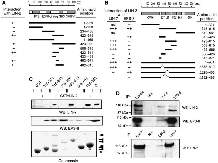
EPS-8 associates with the LIN-2/LIN-7/LIN-10 complex. (A) The indicated fragments of C. elegans EPS-8 were tested in a yeast two-hybrid assay using amino acid 315–615 of C. elegans LIN-2 as bait. (B) The indicated fragments of C. elegans LIN-2 were tested in a yeast two-hybrid assay for interaction with full-length LIN-7 and EPS-8, respectively. Interactions were tested both by His− growth and lacZ activity (+++ strong, ++ medium, + week interaction, defined by lacZ filter staining). (C) GST pull-down assays using the indicated fragments of human LIN-2 CASK cDNA fused to GST and MDCK cell lysates as source of mLIN-7A and mEPS-8 (p97 form). Bound proteins were detected on Western blots with mLIN-7A (upper panel) or mEPS-8 (middle panel) antibodies. The GST::LIN-2 fusion proteins were detected by Coomassie staining (bottom panel). Arrowheads point at GST::LIN-2 fusions, the arrow at GST. (D) Coimmunoprecipitation of mammalian LIN-2 CASK, mEPS-8 and mLIN-7A from MDCK cells. NIS indicates control precipitations with pre-immunoserum. Precipitated proteins were detected on Western blots with polyclonal LIN-2 CASK (upper panel), and monoclonal mEPS-8 antibodies (middle panel). Binding of LIN-2 CASK to mLIN-7A was detected with monoclonal LIN-2 CASK antibodies (lower panel).
