
Figure 2.
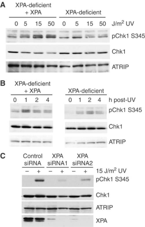
XPA-deficient cells show reduced Chk1 phosphorylation following UV treatment. (A) Isogenic XPA-deficient and XPA-complemented cells were either mock treated or exposed to 0, 5, 15 or 50 J/m2 UV. Cells were harvested after 1 h and lysates were resolved by SDS–PAGE and analyzed by Western blot using phospho-serine 345 Chk1, Chk1 and ATRIP antibodies. (B) XPA-complemented and XPA-deficient cells were mock treated or exposed to 15 J/m2 UV and then harvested at the times shown. Lysates were analyzed as described in panel A. (C) HeLa cells were transfected with control siRNA or one of two siRNAs to XPA and treated with 15 J/m2 UV 48 h later. At 1 h after UV treatment, lysates were prepared and resolved by SDS–PAGE and analyzed by Western blot using phospho-serine 345 Chk1, Chk1, ATRIP and XPA antibodies.