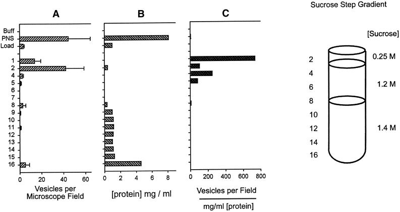
Figure 4.
Sucrose step gradient purification of fluorescent endosomes. The pool from the S200 gel filtration column was made 1.4 M (1.18 g/ml) sucrose and loaded onto the bottom of a sucrose step gradient containing three layers as shown. The gradient was centrifuged to equilibrium, and the fluorescent endosomes (vesicles) (A) floated to the 0.25/1.2 M interface while the bulk of the protein (B) remained at the bottom in fractions 9–16. (C) The specific activity highlights the enrichment of endosomes at this interface. The cloudy layer at fractions 2–4 constituted the final endosome pool and contained the sufficiently high number of endosomes that was required to observe their interaction with microtubules under a microscope.

