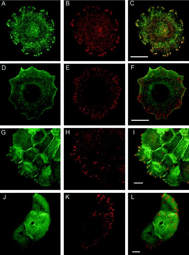
Figure 3.
Double-label confocal immunofluorescence microscopy of SCC4 cells (A–F) and primary keratinocytes (G–L) expressing the wild-type chick β1 subunit (A–C and G–I) or the YPRF mutant (D–F and J–L). Cells were stained with anti-chick β1 antibody (A, D, G and J; green in C, F, I and L) in combination with anti-human β1 antibody (B and E; red in C and F) or anti-vinculin (H and K; red in I and L). Images in C, F, I and L are the merged images of A and B, D and E, G and H, J and K, respectively. Bars, 20 μm.

