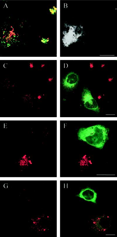
Figure 11.
Dispersed lysosomes in EGFP-Rab7T22N–expressing HeLa cells are defective. In the confocal image pair A-B, a double labeling for Lamp-1 (green) and internalized DiI-LDL (red) is seen. It is evident that internalized LDL is not able to reach the dispersed Lamp-containing lysosomes in the dominant-negative mutant-expressing cell (shown in black and white in the EGFP channel in B). In the confocal image pairs C-D, E-F, and G-H of cells transfected with EGFP-Rab7T22N, the left panels show LysoTracker Red accumulation in lysosomes (red) and the right panels show the merged images with EGFP (green) to identify the mutant-expressing cells and evaluate the degree of expression. Note that the dispersed lysosomes in EGFP-Rab7T22N–expressing cells accumulate little LysoTracker Red (i.e., they are only slightly acidic) compared with the nontransfected cells and that this decreased accumulation reflects the transfection level (G and H). Bars, 20 μm.

