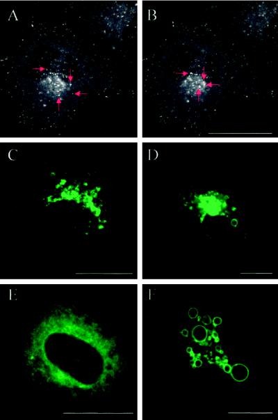
Figure 4.
Detection by confocal microscopy of the EGFP-Rab7 fusion proteins in live HeLa cells. (A and B) Confocal images of a HeLa cell transfected with EGFP-Rab7 wt showing that the fusion protein is associated with vesicular structures that are present throughout the cytoplasm as well as concentrated in a perinuclear aggregate. The images derive from a series of 40 confocal images taken from a live EGFP-Rab7 wt–transfected cell. The red arrows show four examples of fluorescent vesicles moving toward the perinuclear aggregate. (C–F) Confocal images of live HeLa cells expressing EGFP-Rab7 wt (C), the EGFP-tagged active mutant Rab7Q67L at two different expression levels (D and F), and the dominant-negative mutant Rab7T22N (E). The confocal plane and pinhole settings in C, D, and F were chosen to show mainly details of the perinuclear aggregate of structures associated with the EGFP fusion proteins. Note that very high expression levels of the active mutant (F) lead to the formation of large, green fluorescent vacuolar structures. The dominant-negative mutant (E) is distributed throughout the cytoplasm. Bars, 20 μm.

