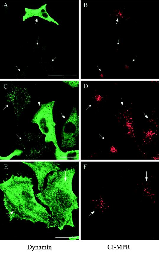
Figure 6.
Expression of mutant dynamin causes redistribution of CI-MPR. HeLa dynK44A cells were grown without tetracycline for 48 h (A–D) or without tetracycline for 72 h and with butyric acid added for the last 24 h (E and F) and double-labeled for dynamin (green) and CI-MPR (red). Note that in the mutant dynamin–expressing cells (large arrows), the CI-MPR signal derives from more distinct and widespread structures than in cells without mutant expression (small arrows). The settings of the confocal microscope were adjusted individually in the two channels to make an optimal distinction between endogenous dynamin and mutant dynamin in each image, as well as an optimal CI-MPR signal. Bar in A, 50 μm; bars in C and E, 20 μm.

