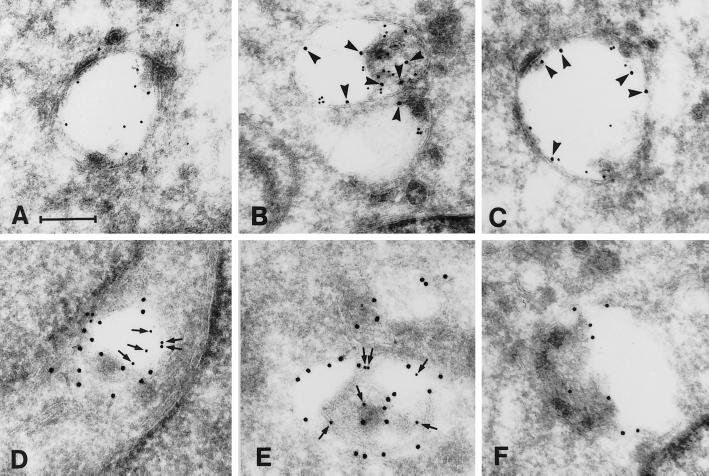
Figure 7.
Representative electron micrographs of the patterns of MPR/Lamp-1 distribution as revealed by immunogold double labeling of ultracryosections. Based on the relative amount of gold labeling for CI-MPR and Lamp-1, endocytic structures were grouped into six types (type 1, MPR+/Lamp-1−; type 2, MPR/Lamp-1 3:1; type 3, MPR/Lamp-1 2:1; type 4, MPR/Lamp-1 1:2; type 5, MPR/Lamp-1 1:3; type 6, MPR−/Lamp-1+). Type 1 (only CI-MPR labeling) represents a classic endosome and type 6 (only Lamp-1 labeling) represents a classic lysosome. A–F show examples of types 1–6, respectively. CI-MPR was detected with 10-nm gold (small arrows in D–F), and Lamp-1 was detected with 15-nm gold (arrowheads in A–C). Bar, 200 nm.

