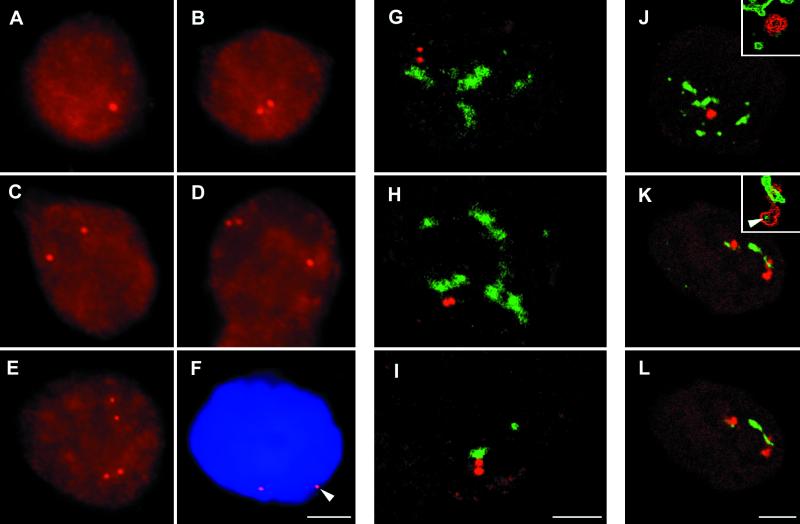
Figure 1.
Organization of EBV genomes (A–F) and relationship between SC35 domains and viral DNA (G–I) or RNA (J–L) in Namalwa cells. The pattern of viral genomes distribution in the interphase nuclei of Namalwa cell has been established by DNA FISH of major internal repeat BamHI W. In 34% of cells, a single dot is observed (A). In 48% of cells, two dots closely spaced within the whole range of ∼3 μm in the x–y plane of each other are observed (B). Seventeen percent of cells have duplicated patterns in three combinations (C–E). A part of the second cell is seen in the bottom part of D. Spatial localization of viral genomes in the interphase nuclei has been performed by means of DNA FISH and DAPI staining. The majority of the signal spatially separated from the nuclear periphery; the minority of genomes have a more peripheral nuclear localization; and some dots are close to nuclear periphery (F, arrowhead). The spatial relationship (documented here by confocal sections) between viral DNA sequences and SC35 domains has been performed by DNA FISH and immunocytochemical mapping by means of the antibody to SC35 splicing factor. In the majority of cells, DNA sequences (red) are observed exclusively outside of the SC35 domains (green; G and I). A smaller fraction of DNA loci, and just one locus in the case of the doublet, is found associated with the outer edge of the SC35 domain (I). Spatial relationship (documented here by confocal sections) between EBV pre-mRNA (red) and SC35 domains (green) has been established by means of RNA FISH and immunocytochemistry. Basically two categories of distribution are observed in the Namalwa cell line. The first category comprises viral RNA spatially separated from SC35 domains (J; the spatial separation is well seen in the inset). The second category includes RNA tightly associated with SC35 domains. This is the major fraction of RNAs forming tracks. RNA signal exhibits various extents of overlap with the SC35 domain (K). The microcluster(s) of SC35 at the pole of RNA accumulations, opposite the SC35 domain, have been observed (K, inset, arrowhead). Insets represent edge-filtered images. K and L are two consecutive confocal sections. Bars, 4.5 μm.

