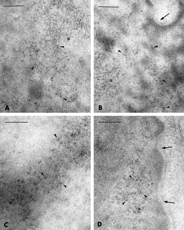
Figure 5.
EM localization of viral RNA in nuclei of Namalwa (A and B) and Raji (C and D) cells. By means of postembedding RNA ISH, most of the visualized RNA (6-nm gold particles; arrowheads) are observed in fibrillogranular structures belonging to PFs. A few gold particles (A, C, and D) are found at the border and/or slightly engulfed in clusters of interchromatin granules (asterisk). Note a more pronounced fine structural heterogeneity of PFs observed in Raji cells than in Namalwa cells. For RNA accumulations found at the outermost nuclear region (B and D), the gold particles are not associated with the nuclear envelope (nuclear pores). Bars, 500 nm.

