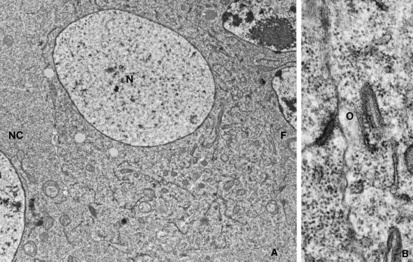
Figure 4.
Wild-type previtellogenic oocyte. (A) Electron micrograph of a stage 6 oocyte, showing the characteristic spherical oocyte nucleus (N), the absence of coated vesicles along the follicle cell (F)–oocyte (O) border (shown at higher magnification in B), and the presence of a dispersed endoplasmic reticulum (arrows), which is absent from nurse cells (NC). Magnification: A, 8100×; B, 42,000×.

