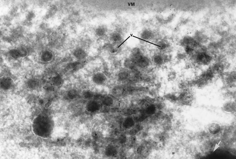
Figure 7.
Ultrastructural localization of Yl at the oocyte cortex. Immunoelectron micrograph of cryothin sections of the cortex of a stage 10 oocyte, stained with 5-nm colloidal gold-labeled secondary antibodies to Yolkless antibody. Antigen is present in coated vesicles (v) and tubules (t), as well as at the surface of nascent yolk platelets (arrow). The vitelline membrane (VM) is at the top. Magnification, 84,000×.

