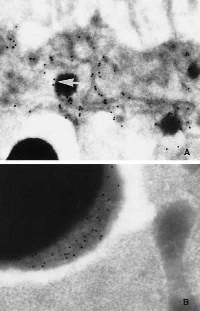
Figure 8.
Ultrastructural localization of Yl. (A) Cryothin section showing the presence of Yolkless in tubules radiating from the surface (top) of a stage 10 oocyte and at the surface of nascent yolk spheres (arrow) (10-nm colloidal gold-labeled secondary antibody). (B) Yolkless antigen is found in the fluffy layer of large yolk granules in a stage 10 oocyte. Magnification: A, 42,000×; B, 42,000×.

