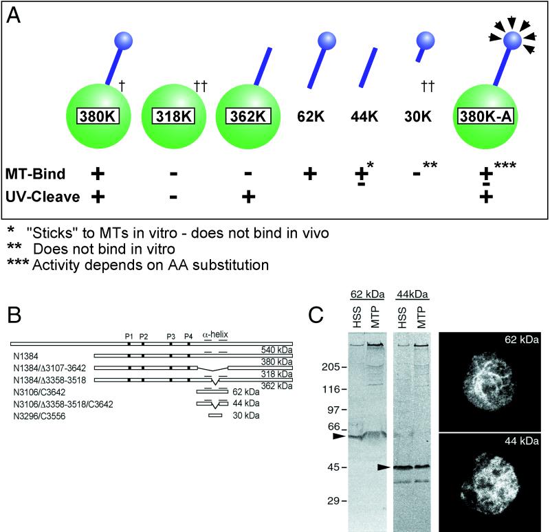
Figure 2.
(A) Summary diagram of microtubule binding for several fragments of the dynein motor domain. The 380K diagrams a complete head; the green represents the motor domain; and the blue represents the stalk and microtubule contact site. The relative positions of these fragments within the DHC are schematically shown in B. †, data from Koonce and Samsó (1996); ††, data from Koonce (1997). (C) In vitro and in vivo microtubule affinity of the 62- and 44-kDa fragments. Both copellet with microtubules as shown on the left immunoblot panels (MTP), probed with the antibody against the 62-kDa fragment. However, only the 62-kDa fragment decorates microtubules in vivo (right). The right panels show two cells fixed and stained with the c-myc antibody, recognizing epitope tags placed on the C terminus of both constructs. Care was taken not to overextract the cells and drive dynein onto the microtubules. Despite the high background, a clear microtubule pattern can be discerned in the 62-kDa–expressing cells, whereas only diffuse cytoplasmic staining can be seen in the 44-kDa–expressing cells.

