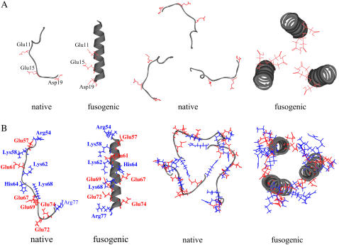
FIGURE 2.
Monomer and trimer structures of the model peptides. The side chain of acidic residue is colored as red, and the side chain of basic residue is colored as blue. (A) The structure of fusion peptide (residues 1–20). The structures of monomer and trimer of the native state are based on the crystal structure (1hgd.pdb). The structure of monomer of the fusogenic state is first generated as a perfect helix and then the trimer structure is generated by maximizing the interaction between three helices with three ionizable residues (Glu-11, Glu-15, and Asp-19) directing inward. (B) The structures of the polypeptide of residues 54–77: The structures of monomer and trimer of the native and fusogenic states are based on the crystal structure (1htm.pdb).

