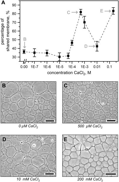
FIGURE 3.
Quantification of Ca2+-induced two-dimensional aggregation of surface-attached giant liposomes composed of 100% eggPC. (A) An image processing algorithm processed the micrographs of giant liposomes after 1 h of flow of CaCl2 to determine the “percentage of shared membrane”, which represents the average percentage of each liposome that is in contact with the visible membrane of adjacent liposomes (N = 3 for all points; error bars represent standard deviations). Images (B–E) show representative phase-contrast micrographs of two-dimensional aggregation after introducing solutions of (B) H2O, (C) 500 μM CaCl2, (D) 10 mM CaCl2, and (E) 200 mM CaCl2 to the liposomes. Note that the liposomes did not exhibit two-dimensional aggregation after introducing an intermediate concentration of 10 mM CaCl2. Scale bars = 75 μm.

