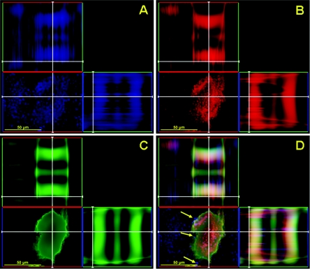
FIG. 2.
Confocal microscopic image of a mature C. neoformans biofilm treated with capsular binding MAb 13F1. Orthogonal images of a mature C. neoformans biofilm show capsular binding MAb 13F1 (blue; GAM-μ-AF) penetration within internal regions of a biofilm (A); metabolically active (red; FUN-1-stained) C. neoformans cells (B); extracellular polysaccharide material (green; ConA-stained) (C); and a superimposition of panels A, B, and C (D). Arrows denote the locations of MAb 13F1 in a mature cryptococcal biofilm. Pictures were taken using a 40× power field. Scale bars, 50 μm.

