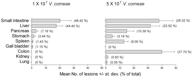
FIG. 2.
Distribution of V. corneae-associated lesions in athymic mice inoculated i.p. with different numbers of organisms. Mice were euthanized after becoming moribund and necropsied for histopathology. The number of lesions with microsporidia were counted at ×10 magnification per tissue section from each athymic mouse, and the means ± standard deviations (st. dev.) of lesions for eight mice per group were plotted. Values presented within parentheses represent the percentage of lesions in each organ compared to the total number of lesions observed in all organs.

