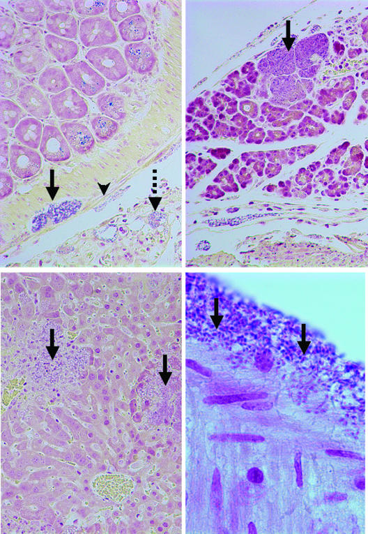
FIG. 3.
Lesions associated with experimental V. corneae infection in athymic mice (Brown Brenn Gram stain). (Top left panel) Small intestine was observed with V. corneae organisms infecting serosal cells (arrowhead), nerve cells of the mesenteric plexus (solid arrow), and cells of the mesentery (dashed arrow) (magnification, ×190). Note that the large zymogen granules in the epithelial cells also stained dark. (Top right panel) Swollen acinar cells of the pancreas were filled with V. corneae organisms (arrow). Inflammation was not usually observed until host cells ruptured (magnification, ×190). (Lower left panel) V. corneae organisms were observed in the liver and lysed random groups of hepatocytes (arrows). Necrotic foci contained debris, neutrophils, and V. corneae organisms (magnification, ×190). (Lower right panel) The light-staining posterior vacuoles of dark-staining V. corneae organisms in intestinal serosa can be observed (arrows) (magnification, ×950).

