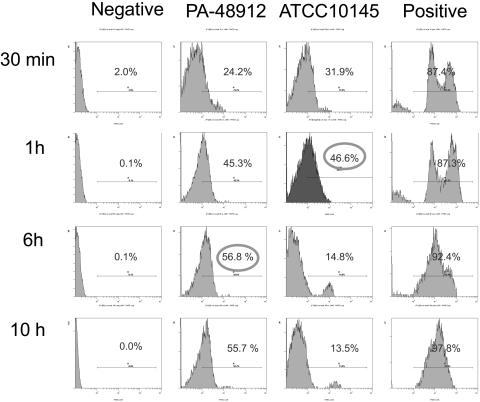
FIG. 3.
Flow cytometric analysis of liposome-bacterium interactions. A sensitive laboratory strain, ATCC 10145, and a resistant clinical strain, PA-48912-2, were incubated with PBS (left panels), labeled empty liposomes (middle panels), or free PKH2-GL (right panels) for 0.5, 1, 6, and 10 h at 37°C with agitation. Percentages of the labeled bacterial cells are indicated.

