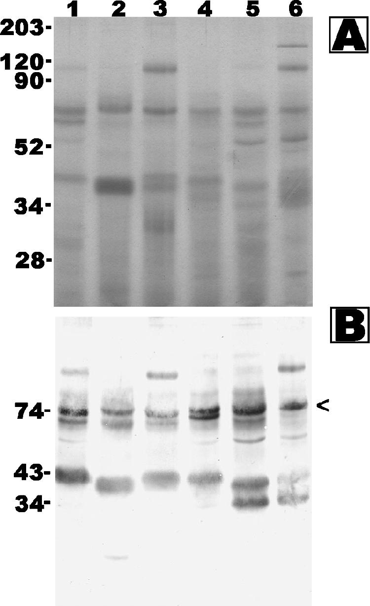
FIG. 3.

Coomassie brilliant blue-stained SDS-PAGE gel (A) and Western blot analysis (B) of Pythium insidiosum CFAs P1 (lane 1), P15 (lane 2), P10 (lane 3), P6 (lane 4), P8 (lane 5), and P7 (lane 6) probed with S12 pythiosis serum. Molecular markers (A) or predicted sizes (B) are shown in kDa on the left.
